Extraoral radiograph Panoramic X-ray Tomograms Cephalometric projections Sialography Computed tomography 10. A periapical x-ray or PA film will show one or two teeth in their entirety in one single image right from the crown of the tooth which is the part exposed in the mouth to the very tips of the tooth roots located in the jawbone as well as.

Periapical Radiography Pocket Dentistry
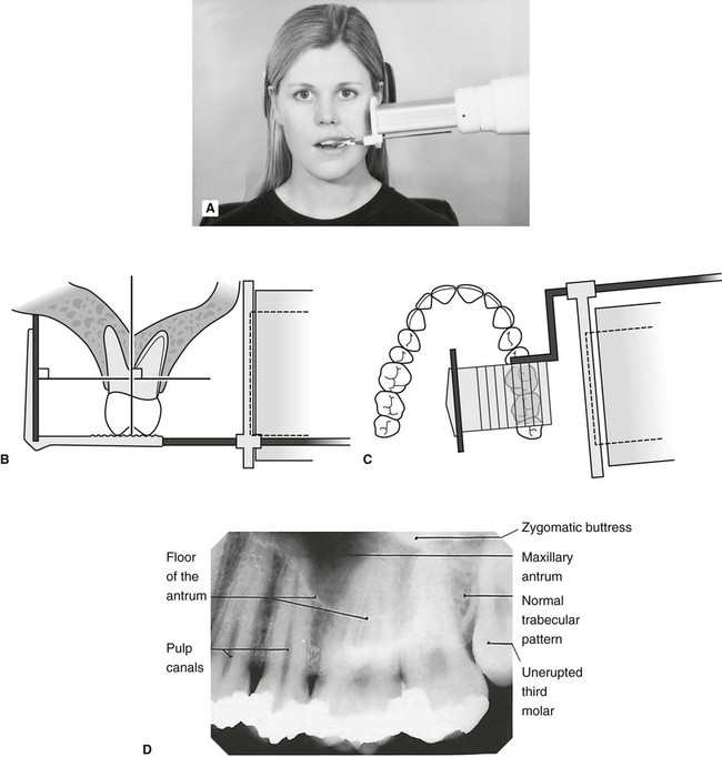
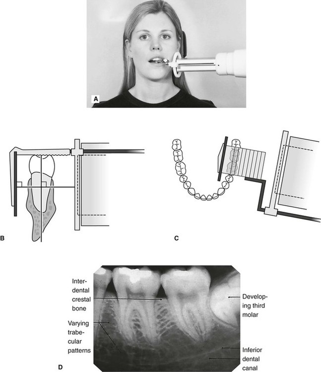
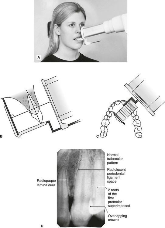
BISECTING SHORT-CONE PERIAPICAL EXPOSURE TECHNIQUES.
. Ad Publishing Open Access Peer Reviewed Research related to the field of Scanning. Our Service Includes Free Proofreading Language Editing. Intraoral periapical radiographs can be produced using two different techniques.
To take a periapical exposure the hygienist or x-ray technician places a small photosensitive imaging plate coated with phosphorus into a sterile wrapper and inserts it into the patients mouth just like a conventional X-ray film card. The film is placed parallel to the long axis of the tooth in question and the central x-ray beam should be directed perpendicular to the long axis of the tooth. The target-film distance is 8 inches.
These patients may include adults with low palatal vaults and children. The X-ray head is directed at right angles vertically and horizontally of both the tooth and the image receptor. Both techniques have advantages and disadvantages.
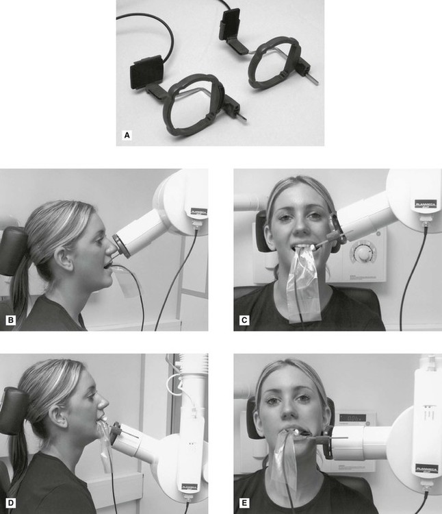
The paralleling technique results in good quality x-rays with a minimum of distortion and is the most reliable technique for taking periapical x-rays. The X-ray tubehead is then aimed at right angles vertically and horizontally to both the tooth and the image. The periapical film is held between the incisor teeth as if it were an occlusal film for all anterior periapical radiographs.
The extraoral periapical radiographic technique was performed for both maxillary and mandibular teeth using Newman and Friedman technique2. Periapical Lesion Diagnosis Support System Based on X-ray Images Using Machine Learning Technique. The bisecting-the-angle technique and the more commonly used long cone paralleling technique.
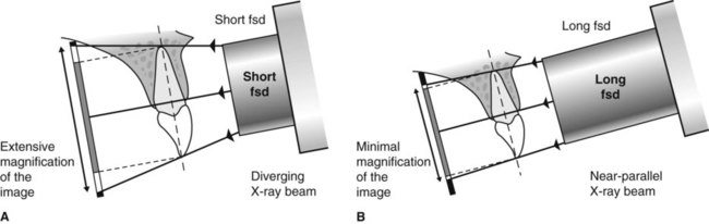
Periapical X-rays are used to detect any abnormalities of the root structure and surrounding bone structure. Vo TN Ngoc 1 Do H Viet 2 Le K Anh 3 Dinh Q Minh 4 Le L Nghia 5 Hoang K Loan 6 Tran M Tuan 7 Tran T Ngan 8 Nguyen T Tra 9. The resulting image x-ray is somewhat larger using the short cone rather than using a long cone see figure 4-1.
A long cone is used to take x-rays with paralleling exposure techniques. The central ray is directed to pass at a perpendicular angle to both the tooth and the film. Parallel technique The image receptor is placed in a holder and placed in the mouth parallel to the longitudinal axis of the tooth under.
The image receptor is placed in a holder and positioned in the mouth parallel to the long axis of the tooth under. Periapical x ray techniques Written By weisinger Saturday March 12 2022 Add Comment Edit. The technique involves a constant x-ray cone position which is perpendicular to the floor for maxillary incisors and parallel to the floor for mandibular incisors.
The patient was positioned upright with hisher mouth was opened as wide as possible to allow the X-ray beam to pass to the sensor unobstructed from the opposite side of the mouth. The bisecting technique may have to be used for patients unable to accommodate the film positioningdevice used in the paralleling technique. Pericoronal refers to the crown area of the tooth.
Occlusal X-rays show full tooth development and placement 9. Periapical radiography is a commonly used intraoral imaging technique in radiology and may be a component of your radiologic examination. 1 Department of Pediatric Dentistry School of Odonto-stomatology Hanoi Medical University Dong Da Hanoi Vietnam 26 Department of.
The bisecting short-cone and paralleling long-cone techniques are two of the most commonly used techniques. Single periapical radiographs are often made of individual teeth or groups of teeth to obtain information for treatment or diagnosis of localized diseases or abnormalities. The dental assistant works with the dentist in providing patient treatment including restorations x-rays and preventive services.
By using a filmsensor holder with fixed image receptor and. Since the slope and curvature of the dental arches and the alveolar processes will not permit the film to be held close to the teeth. Periapical X-rays show the entire tooth from the exposed crown to the end of the root and the bones that support the tooth.
There are two types of techniques used for periapical radiographs. By using a film sensor holder with still. Bisecting angle and paralleling.
The Long Cone Paralleling Technique. Submit Your Paper With Hindawi. Tracings from lateral skull radiographs taken of dried skulls of various ages were used to.
The sensor was placed on the. Most frequently used radiography is for the periapical which is performed by the bisecting Thus when considering the execution of the radiographic technique and the possibility of errors that occur during the exposure of X-ray image XR receptors it is important to identify those that occur more frequently. Disadvantages to the bisecting technique.
The bisecting plane is halfway between the plane of the dental film and the. A short cone is used to take x-rays with bisecting angle exposure techniques. Read customer reviews find best sellers.
Ad Browse discover thousands of brands. The X-ray is taken and the exposed plate is then loaded into a scanner or processor which reads the image. Periapical film is held parallel to the long axis of the tooth using film-holding instruments.
Periapical X-rays. Periapical radiographs provide important information about the teeth and surrounding bone. These X-rays are used to find dental problems below the gum line or in the jaw such as impacted teeth tooth fractures abscesses tumours and bone changes linked to some diseases.

How Make Periapical X Ray
Periapical Radiography Pocket Dentistry

Periapical Radiography Pocket Dentistry
Periapical Radiography Clinical Gate
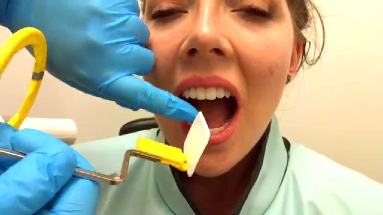
How To Take Periapical Radiographs Youtube
Periapical Radiography Clinical Gate
Periapical Radiography Clinical Gate
Periapical Radiography Clinical Gate
0 comments
Post a Comment